

Investigating stress-induced mitochondrial dysfunction and its role in systemic health
Metabolic stress is a common occurrence for active-duty military service members. It occurs during periods of sustained physiologic challenges such as continuous physical activity, thermal stress, severe caloric deficits and sleep deprivation, which warfighters routinely experience. These stressors, both on their own and in combination, eventually lead to metabolic dysfunction, which can degrade a warfighter’s operational performance.
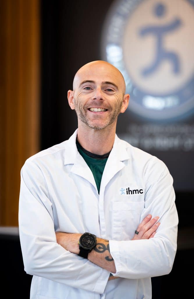
HMC Research Scientist Zachary Graham has a new three-year Air Force Office of Scientific Research grant to gain a better understanding of stress-induced mitochondrial dysfunction. Mitochondria are specialized, double-membrane structures within cells that serve as the primary source of energy production. They are often referred to as the powerhouses of the cell.
In addition to regulating energy metabolism, mitochondria also regulate controlled cell death, called apoptosis. They also can drive a cellular pro-inflammatory environment when normal function is compromised. Dysfunctional mitochondria create excessive reactive oxygen and nitrogen species (ROS/RNS) that can damage proteins, lipids and DNA/RNA; these changes also activate inflammation. Gaining a better understanding of mitochondria and their role in stress-induced mitochondrial dysfunction can provide critical insights into the sources of cellular energy as well as breakdowns of cellular energy.
Graham, who has been at IHMC for the past five years, primarily researches how exercise can be implemented to improve the health and quality of life of people across disease states and lifespan. His projects focus on in-depth phenotyping, including cognitive and physical performance, fresh tissue and in vitro mitochondrial function, and high-resolution integrative molecular multiomics profiling.
Below is a conversation with Graham about his research and his three-year grant to study the role of stress-induced mitochondrial dysfunction on the subcellular metabolome.
Much of your research over the years has focused on mitochondria, which are membrane-bound organelles that are often referred to as the powerhouses of the cell because they produce the energy that’s critical for cellular functions. What led you to first start researching mitochondrial dysfunction?
I first started about 12 to 13 years ago during my postdoc. I was really interested in first, how to protect skeletal muscle mass after paralysis, which we induced using a spinal cord injury in mice. There are pharmaceuticals that exist that target key molecules that reduce muscle loss and we tested a bunch of them with little success in animals. Spinal cord injury is not a static injury and a lot of people recover some function in early weeks during recovery as the swelling around the spinal cord reduces. I was interested in finding a way to slow the loss of muscle during this recovery period as we thought this would allow someone to have more muscle when they start recovery, which we believed would help a person recover faster.
I then started to become interested in mitochondrial function, which makes up a lot of our muscle. If we couldn’t preserve muscle mass, maybe at least we could preserve muscle metabolic function. Some 5-15 percent of our muscle cells are mitochondria, depending on the fiber type and there are drugs that exist that are close to market or at market that you can use to target mitochondria and importantly, you can inject into a human. My general thought is that you’re only as healthy as your muscle, and your muscle is only as healthy as your muscle mitochondria. That’s kind of how I think about it.
In the abstract for this three-year grant you’re working on, you write that the study is designed to better understand the role of stressed- induced mitochondrial dysfunction. What is the question you’re attempting to answer with this study?
We have what we refer to as transient stress, which is a good thing. Exercise is the best example of this. A transient stress that demands everything from your body provides a molecular and cellular response to build a reserve against that stress in the future. So, if you’ve never exercised and you start to exercise for a while, you get sore. But after exercising for a little while, you’re not sore. That’s in response to that stress. It’s also why if you don’t exercise for a bit, you’re slower or weaker; your body recognizes you no longer need that reserve and gets rid of it.
But then take somebody who goes through sustained, chronic stress with little to no rest. For example, you take a high-level warfighter on a three-week operation who is carrying
a 60-pound ruck for 10 hours in a hot, mountainous region, not sleeping and not eating and is psychologically stressed. Stress is cumulative. Even as single-factor stresses, all of those things reduce the body’s ability to perform normally. But because stress is cumulative, all of these factors add on to each other.
So, taking exercise again as an example, you take three years of exercising correctly to train for the Olympics. Your goal is to peak at a specific date and do better than you’ve ever done. Leading up to the competition you train a little bit more than normal. Then you get anxious so you’re sleeping and eating less and you’re not giving your body a chance to rest and recover. If you continue on this path, your performance will be worse. That’s a simplified version and in the exercise world that’s what we refer to as overtraining but can easily be translated to any activity or profession that stresses the body.
Overtraining, or overuse, isn’t unique to warfighters and other active-duty service members. Elite athletes also experience periods of sustained physiologic challenges that lead to overtraining and overuse.
Yes, military operators and various types of athletes, both recreational and competitive, are always stressed. And in terms of systemic function, this stress affects the entire body, not just one part of the body, like the lungs. And even though military operators and athletes are elite for a reason, nobody is impenetrable to overuse or overtraining.
Take an elite cyclist. They are in better shape than 99.99 percent of people even on a bad day. But if they’re racing the Tour de France and they overtrain too long without giving their body time to rest and recover, their performance compared to their peers in the race is going to be worse. Military operators, however, face a different scenario. They may not be in a situation that is voluntary and orders may place them in rugged terrain in bad weather where they have a ruck and have to hike up a mountain while carrying their weapons and all of their food while being shot at. Their training allows them to manage all these stresses for a day or two and maintain high levels of performance. Some people can handle it longer than others, but everybody has a breaking point and the cure is rest and recovery.
So how is the study being designed?
It’s all in vitro, so it’s all cells – no human participants. The benefit of in vitro study is that you don’t need institutional approval to protect your participants or your mice. You just buy the cells and you get them up and going. This helps with scientific rigor and repeatability because other people can buy the same exact cells and replicate what you’re doing and what you have found. Then you just start cranking through a bunch of experiments.
What are you hoping to find?
What I’m interested in is how the cells themselves communicate stress. Our cells release what are called extracellular vesicles (EVs), which are small membrane-bound particles released by cells and play crucial roles in intercellular communication by carrying proteins, nucleic acids, and other biomolecules between cells. I believe almost all cells can
secrete EVs and it’s a way for our cells to talk through our bodies and system. Within the framework of the grant, we’re going to look at how these cells respond to stress. For a few examples, we will see the timecourse of stress and how fast our cells start to change the EV cargo. We think mitochondrial stress is a major regulator of this and the cells will actually package damaged mitochondria into these EVs. We will also collect the EVs from stressed cells and then put them in healthy cells and see how the healthy cells react. By using cell culture models, we can test all of this in a rigorous way where we don’t have to inject unknown things into a human, but also provide early translational directions where we may identify something we could target with a drug.
Latest News
- Investigating stress-induced mitochondrial dysfunction and its role in systemic health
- IHMC studies the potential of exoskeletons to reduce the biomechanical injuries shipyard workers face
- A Q&A With IHMC’s new CEO, Dr. Timothy Broderick
- Centering the Human in AI Decision Making
- Investigating exercise’s potential to reverse hallmarks of aging
- Alex wins Army’s xTechHumanoid competition
- STEM-Talk: Is AI out of control? Could it kill humanity? Ken Ford discusses the real and imagined risks of AI
- STEM-Talk: Ken and Dawn tackle listener questions on protein, genetics, resistance training, Moon landings and more
- STEM-Talk: Ken Ford separates what’s real in AI from hype and science fiction